Anatomical Structures
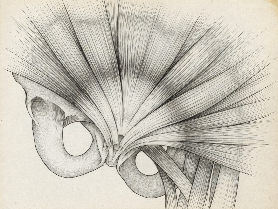
The Muscular Sling
Levator Ani Complex
The levator ani is the primary muscle group of the pelvic floor, consisting of the pubococcygeus, puborectalis, and iliococcygeus. During a vaginal delivery, these muscles undergo extreme stretching, sometimes elongating up to 300% of their resting length. Micro-tears or avulsions here are the primary cause of postpartum weakness.
Endopelvic Fascia
Connective tissue acts as the suspension bridge for pelvic organs. The uterosacral and cardinal ligaments suspend the uterus and upper vagina. Hormonal changes (specifically relaxin) during pregnancy soften these structures, requiring careful load management postpartum to allow for tissue recoiling.
Organ Placement
The Anterior, Middle, and Posterior Compartments
Clinically, the pelvis is divided into three compartments. The anterior holds the bladder and urethra. The middle contains the uterus and vagina. The posterior houses the rectum. Understanding these compartments is vital for diagnosing specific types of prolapse (cystocele, apical prolapse, rectocele).